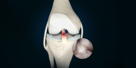
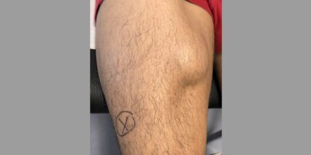
The incidence of chondral and osteochondral defects is increasing due to the raised activity profile of the population and modern, continually improving MRI diagnostic methods. The incidence of cartilaginous defects in the knee among athletes is given as up to 36 % [13].
Untreated lesions cause higher mechanical loading of the surrounding intact cartilage [9, 16, 18] and have an effect on the subchondral bone [27] and on the intra-artikular mlieu, with an increase of cytokine concentration [14] and thus a premature onset of osteoarthritis. This not only limits the patients’ function, but also causes considerable costs to the public health system. Thus, suitable cartilage reconstruction techniques with the specific regeneration of hyaline- or hyaline-like cartilage are required. A number of different surgical procedures are already available for the treatment of focal chondral lesions, including techniques like bone marrow stimulation (microfracture (MFx), autologous matrix-induced chondrogenesis (AMIC)), osteochondral auto- or allograft transplantation surgery (OATS), and autologous chondrocyte implantation (ACI) [5, 11 – 13, 33]. Since each of these techniques has pros and cons, the treatment of chondral lesions has not been standardised and remains a challenge.
ACI is currently seen as the treatment of choice for moderate to large chondral defects, since this procedure leads to hyaline- or hyaline-like cartilage substance with good long-term clinical outcomes [6, 7, 15, 17, 28]. The disadvantages of ACI are the high laboratory costs for cell expansion, limited availability in some cases, and the need for two-step surgical procedures [5, 12, 32]. In order to overcome these disadvantages, one-step procedures such as the implantation of fragmented autologous or allogenic cartilage have been developed (Minced Cartilage Implantation (MCI)). The underlying principle was already described in the early 1980s by Albrecht et al. [1, 2] and picked up again by Lu et al. in 2006 [22]. Over the past few years interest in MCI has grown considerably especially due to several advantages, such as being a single stage procedure that can be performed in an arthroscopic or mini-arthrotomy surgical approach and may offer strong biologic potential [5, 32].
Biology
In their in vivo milieu chondrocytes have the potential to proliferate physiologically [3, 32]. Furthermore, mechanical stimuli play an important role in chondrocyte proliferation and chondrogenic differentiation [36, 37]. This complex biochemical and biomechanical intra-articular milieu, that can barely be reproduced in vitro, could be a major advantage regarding the biological potential of the MCI procedure. It has been shown that mincing healthy cartilage “activates” the chondrocytes and leads to a physiological reaction with chondrogenic proliferation and the production of extracellular matrix (ECM) [22 – 24, 32]. Mincing can be achieved with a scalpel, specially developed mincing devices, or arthroscopic shavers [19, 20, 32]. The cartilage is harvested from the margins of the chondral defect or from zones of the joint that are subjected to less loading. The outgrowth of activated chondrycytes is promoted by this enlargement of the tissue surface [4, 5, 20, 22, 32]. Ultimately this leads to the regeneration of hyaline and/or hyaline-like cartilage [22, 32, 35].
Surgical technique
The preoperative planning of a chondroplasty procedure includes a mandatory MRI and conventional radiographs (whole-leg) [31] to detect and treat any comorbidities such as ligamentous instability, meniscus defects or mechanical axial malalignment. The final planning of the chondroplasty is only completed following detailed arthroscopic diagnostic investigation of the defect. The cartilage can then be harvested with osteochondral cylinders from zones that are barely load-bearing (e.g. the intercondylar notch), or using ring curettes and shavers [31, 33]. When using osteochondral cylinders the cartilage must be separated from the bone and then minced with a scalpel or shaver until it has reached a paste-like consistency. During arthroscopic cartilage harvesting this is done exclusively with a shaver [33]. After preparing the defect and creating stable cartilage margins the joint is aspirated, the defect zone is dried, and the minced cartilage is introduced into the defect. Depending on the technique being used, autologous thrombin and PRP, fibrin glue and/or a membrane are used for stable fixation of the fragments [25, 26, 31 – 33].
Clinical data
Clinical evidence on autologous MCI is still limited [8, 10, 25]. In 2015, Christensen et al. [8] treated eight patients with osteochondrosis dissecans of the knee joint with a combination of autologous bone graft and autologous cartilage fragments embedded in fibrin glue (autologous dual-tissue transplantation (ADTT)). One year later there was a marked improvement in the MOCART score (Magnetic Resonance Observation of Cartilage Repair Tissue) from 22 to 52 points. In 2019, Massen et al. [25] conducted a consecutive two-year study of patients with (osteo-)chondral lesions who had been treated with autologous MCI. At the final follow-up examination a significant reduction in pain was observed. Moreover, a significant radiological improvement in the MOCART score was seen. In 2020, Cugat et al. [10] treated 15 patients with full-surface (osteo-)chondral lesions using autologous MCI embedded in platelet-poor plasma (PPP) and PRP. After 15 months they also observed statistically significantly better scores on the visual analogue scale (VAS) for pain, the Lysholm score, the subjective International Knee Documentation Committee (IKDC) score, the Western Ontario and the McMaster Universities Osteoarthritis Index (WOMAC) for pain and function, the Lequesne-Index and the Short Form 12 (SF-12). While the above-named clinical studies were conducted on the knee joint, autologous MCI is also used in other joints (hip, shoulder, ankle) [21, 29, 30, 34]. In summary it may be said that the clinical data to date show good results with low complication and revision rates which are comparable to other cartilage repair techniques (ACI).
Case study
A 37-year-old patient, an active sportsman, consulted us after multiple left knee sprains; first sprain in 2005. Since then intermittent symptoms in the left knee joint, prone to swell. The clinical examination showed articular effusion with pain on pressure over the medial joint space and mild crepitation in the medial compartment when testing movement, ROM extension/flexion 3-0-145° pain-free. Joint with stable ligaments. The Knee Injury and Osteoarthritis Outcome Score (KOOS) for pain was 50 before surgery, the quality of life score for the knee was 25 points, and 44 for activities of daily living (0 = extreme knee problems, 100 = no knee-related impairment). The Marx activity rating scale (MARS) was initially 0 points (0 = lowest physical and sporting activity, 16 = highest physical and sporting activity). The MRI showed grade 4 cartilage damage over the medial femoral condyle (Fig. 1). The preoperative AMADEUS score for the medial cartilage lesion was 60 points. Minced cartilage implantation was indicated.
The arthroscopic operation with the implantation of minced cartilage in the medial femoral condyle using the product Autocart (Arthrex) was performed. During the operation we diagnosed a 3 cm² ICRS grade 3B chondral lesion over the medial femoral condyle (Fig. 2). The postoperative course was complication-free. Follow-up treatment consisted of six weeks’ partial 15 kg weight-bearing on the left. The range of motion was limited to 60° for weeks one, two and three, and to 90° for weeks four, five and six. The patient was initially splinted with a Mecron brace, which was replaced with a rigid frame brace in the later course. He had physiotherapy for three months after surgery. At the check-up two months after surgery he still had some residual symptoms with a ROM in extension/flexion of 0-0-90° and his quadriceps muscles were still weakened.

 Fig. 2 Intra-operative arthroscopic images showing the untreated cartilage damage (a)
and the lesion after preparation of stable cartilage margins (b) and implantation of the minced cartilage (c).
After eight months the patient then reported a satisfactory surgical outcome with improved movement and a clinically irritation-free knee joint. One year after surgery the patient was still satisfied and was able to re-initiate low-impact sporting activities (cycling). After two years the patient had reached his regular everyday level, and intensification of sporting activities in the low-impact range was possible. The clinical outcome parameters showed an improvement in the KOOS score for pain (from 50 to 53 points), knee-related quality of life (from 25 to 38 points) and activities of daily living (from 44 to 71 points). The MARS had also increased from 0 points before the operation to 6 points afterwards. The MRI two years after surgery showed a corresponding satisfactory outcome with a MOCART score of 95 points (Fig. 3).

Summary and outlook
On the basis of the available in vitro and in vivo data, autologous MCI is a promising one-step chondral repair procedure with great biological and clinical potential. Further medium- to long-term comparative studies on large patient cohorts with clinical, functional and radiological data are required to determine the optimal defect size for MCI and the durability of the repair cartilage, and to enable comparison with other, established chondral repair procedures.
Literature
- Albrecht F, Roessner A, Zimmermann E (1983) Closure of osteochondral lesions using chondral fragments and fibrin adhesive. Arch Orthop Trauma Surg (1978) 101:213-217
- Albrecht FH (1983) [Closure of joint cartilage defects using cartilage fragments and fibrin glue]. Fortschr Med 101:1650-1652
- Barry F, Murphy M (2013) Mesenchymal stem cells in joint disease and repair. Nat Rev Rheumatol 9:584-594
- Bonasia DE, Marmotti A, Mattia S, Cosentino A, Spolaore S, Governale G, et al. (2015) The Degree of Chondral Fragmentation Affects Extracellular Matrix Production in Cartilage Autograft Implantation: An In Vitro Study. Arthroscopy 31:2335-2341
- Bonasia DE, Marmotti A, Rosso F, Collo G, Rossi R (2015) Use of chondral fragments for one stage cartilage repair: A systematic review. World J Orthop 6:1006-1011
- Brittberg M, Lindahl A, Nilsson A, Ohlsson C, Isaksson O, Peterson L (1994) Treatment of deep cartilage defects in the knee with autologous chondrocyte transplantation. N Engl J Med 331:889-895
- Chimutengwende-Gordon M, Donaldson J, Bentley G (2020) Current solutions for the treatment of chronic articular cartilage defects in the knee. EFORT Open Rev 5:156-163
- Christensen BB, Foldager CB, Jensen J, Lind M (2015) Autologous Dual-Tissue Transplantation for Osteochondral Repair: Early Clinical and Radiological Results. Cartilage 6:166-173
- Coetzee JC, Giza E, Schon LC, Berlet GC, Neufeld S, Stone RM, et al. (2013) Treatment of osteochondral lesions of the talus with particulated juvenile cartilage. Foot Ankle Int 34:1205-1211
- Cugat R, Alentorn-Geli E, Navarro J, Cusco X, Steinbacher G, Seijas R, et al. (2020) A novel autologous-made matrix using hyaline cartilage chips and platelet-rich growth factors for the treatment of full-thickness cartilage or osteochondral defects: Preliminary results. J Orthop Surg (Hong Kong) 28:2309499019887547
- Dekker TJ, Aman ZS, DePhillipo NN, Dickens JF, Anz AW, LaPrade RF (2021) Chondral Lesions of the Knee: An Evidence-Based Approach. J Bone Joint Surg Am 103:629-645
- Evenbratt H, Andreasson L, Bicknell V, Brittberg M, Mobini R, Simonsson S (2022) Insights into the present and future of cartilage regeneration and joint repair. Cell Regen 11:3
- Flanigan DC, Harris JD, Trinh TQ, Siston RA, Brophy RH (2010) Prevalence of chondral defects in athletes‘ knees: a systematic review. Med Sci Sports Exerc 42:1795-1801
- Fraser A, Fearon U, Billinghurst RC, Ionescu M, Reece R, Barwick T, et al. (2003) Turnover of type II collagen and aggrecan in cartilage matrix at the onset of inflammatory arthritis in humans: relationship to mediators of systemic and local inflammation. Arthritis Rheum 48:3085-3095
- Gikas PD, Bayliss L, Bentley G, Briggs TW (2009) An overview of autologous chondrocyte implantation. J Bone Joint Surg Br 91:997-1006
- Gratz KR, Wong BL, Bae WC, Sah RL (2009) The effects of focal articular defects on cartilage contact mechanics. J Orthop Res 27:584-592
- Grevenstein D, Mamilos A, Schmitt VH, Niedermair T, Wagner W, Kirkpatrick CJ, et al. (2021) Excellent histological results in terms of articular cartilage regeneration after spheroid-based autologous chondrocyte implantation (ACI). Knee Surg Sports Traumatol Arthrosc 29:417-421
- Guettler JH, Demetropoulos CK, Yang KH, Jurist KA (2004) Osteochondral defects in the human knee: influence of defect size on cartilage rim stress and load redistribution to surrounding cartilage. Am J Sports Med 32:1451-1458
- Hunziker EB, Quinn TM, Hauselmann HJ (2002) Quantitative structural organization of normal adult human articular cartilage. Osteoarthritis Cartilage 10:564-572
- Levinson C, Cavalli E, Sindi DM, Kessel B, Zenobi-Wong M, Preiss S, et al. (2019) Chondrocytes From Device-Minced Articular Cartilage Show Potent Outgrowth Into Fibrin and Collagen Hydrogels. Orthop J Sports Med 7:2325967119867618
- Lorenz CJ, Freislederer F, Salzmann GM, Scheibel M (2021) Minced Cartilage Procedure for One-Stage Arthroscopic Repair of Chondral Defects at the Glenohumeral Joint. Arthrosc Tech 10:e1677-e1684
- Lu Y, Dhanaraj S, Wang Z, Bradley DM, Bowman SM, Cole BJ, et al. (2006) Minced cartilage without cell culture serves as an effective intraoperative cell source for cartilage repair. J Orthop Res 24:1261-1270
- Marmotti A, Bruzzone M, Bonasia DE, Castoldi F, Rossi R, Piras L, et al. (2012) One-step osteochondral repair with cartilage fragments in a composite scaffold. Knee Surg Sports Traumatol Arthrosc 20:2590-2601
- Marmotti A, Bruzzone M, Bonasia DE, Castoldi F, Von Degerfeld MM, Bignardi C, et al. (2013) Autologous cartilage fragments in a composite scaffold for one stage osteochondral repair in a goat model. Eur Cell Mater 26:15-31; discussion 31-12
- Massen FK, Inauen CR, Harder LP, Runer A, Preiss S, Salzmann GM (2019) One-Step Autologous Minced Cartilage Procedure for the Treatment of Knee Joint Chondral and Osteochondral Lesions: A Series of 27 Patients With 2-Year Follow-up. Orthop J Sports Med 7:2325967119853773
- Matsushita R, Nakasa T, Ishikawa M, Tsuyuguchi Y, Matsubara N, Miyaki S, et al. (2019) Repair of an Osteochondral Defect With Minced Cartilage Embedded in Atelocollagen Gel: A Rabbit Model. Am J Sports Med 47:2216-2224
- Minas T, Nehrer S (1997) Current concepts in the treatment of articular cartilage defects. Orthopedics 20:525-538
- Riboh JC, Cvetanovich GL, Cole BJ, Yanke AB (2017) Comparative efficacy of cartilage repair procedures in the knee: a network meta-analysis. Knee Surg Sports Traumatol Arthrosc 25:3786-3799
- Roth KE, Klos K, Simons P, Ossendorff R, Drees P, Maier GS, et al. (2021) [Cartilage chip transplantation for cartilage defects of the first metatarsophalangeal joint]. Oper Orthop Traumatol 33:480-486
- Roth KE, Ossendorff R, Klos K, Simons P, Drees P, Salzmann GM (2021) Arthroscopic Minced Cartilage Implantation for Chondral Lesions at the Talus: A Technical Note. Arthrosc Tech 10:e1149-e1154
- Salzmann GM, Calek AK, Preiss S (2017) Second-Generation Autologous Minced Cartilage Repair Technique. Arthrosc Tech 6:e127-e131
- Salzmann GM, Ossendorff R, Gilat R, Cole BJ (2021) Autologous Minced Cartilage Implantation for Treatment of Chondral and Osteochondral Lesions in the Knee Joint: An Overview. Cartilage 13:1124S-1136S
- Schneider S, Ossendorff R, Holz J, Salzmann GM (2021) Arthroscopic Minced Cartilage Implantation (MCI): A Technical Note. Arthrosc Tech 10:e97-e101
- Schumann J, Salzmann G, Leunig M, Rudiger H (2021) Minced Cartilage Implantation for a Cystic Defect on the Femoral Head-Technical Note. Arthrosc Tech 10:e2331-e2336
- Tseng TH, Jiang CC, Lan HH, Chen CN, Chiang H (2020) The five year outcome of a clinical feasibility study using a biphasic construct with minced autologous cartilage to repair osteochondral defects in the knee. Int Orthop 44:1745-1754
- Tsuyuguchi Y, Nakasa T, Ishikawa M, Miyaki S, Matsushita R, Kanemitsu M, et al. (2021) The Benefit of Minced Cartilage Over Isolated Chondrocytes in Atelocollagen Gel on Chondrocyte Proliferation and Migration. Cartilage 12:93-101
- Wang N, Grad S, Stoddart MJ, Niemeyer P, Reising K, Schmal H, et al. (2014) Particulate cartilage under bioreactor-induced compression and shear. Int Orthop 38:1105-1111
Autoren
ist Assistenzarzt Hüft- und Kniechirurgie an der Schulthess Klinik Zürich.
Seine Forschungstätigkeit liegt in der Hüft- und Kniechirurgie, Gelenkserhalt und Gelenksrekonstruktion. Sein Schwerpunkt in der Sportorthopädie/-traumatologie ist die Knorpelrekonstruktion.
ist Facharzt für Orthopädie und Unfallchirurgie. Er hat seinen Lehrauftrag an der medizinischen Fakultät der Universität Freiburg im Breisgau. Sein Fachgebiet ist die Kniechirurgie. Er ist Partner am Gelenkzentrum Rhein-Main und Leitender Oberarzt Sektion Kniechirurgie an der Schulthess Klinik in Zürich. Professor Salzmann ist AGA Instruktor und beratender Arzt der Eintracht Frankfurt, Fußball.
ist Assistenzarzt für Unfallchirurgie und Orthopädie an der Sektion Sportorthopädie, Klinikum Rechts der Isar, TU München. Außerdem betreut er die HC Innsbruck – Die Haie (Eishockey) sowie die Italienische Faustball Nationalmannschaft.


