")
REMS (Radiofrequency Echographic Multi Spectrometry) technology is an innovative, ultrasound-based method for assessing bone mineral density and bone structure that can be used to screen bone health and support the diagnosis and monitoring of osteopenia and osteoporosis [1]. Unlike conventional DXA measurement (dual energy X-ray absorptiometry), which has been considered the gold standard in osteoporosis diagnostics to date, REMS is based on the analysis of unfiltered ultrasound data, which is evaluated using special algorithms for spectral analysis, allowing not only bone mineral density (BMD) but also the structural quality of the bone to
be assessed [1, 2].
For each scan line, spectra are generated from these spectral signatures, which are then compared with reference data from healthy and osteoporotic bones [1]. The BMD values calculated from this (in g/cm2) are then converted into T and Z scores using linear transformations [1]. In comparison, conventional ultrasound methods are usually based solely on pure image information (B-mode) [1]. Devices based on this technology, such as the models developed by Echolight, enable precise, fast, and reproducible measurements directly on the lumbar spine (L1 – L4) and femoral neck – analogous to the DXA regions. A small ultrasound probe is placed on the skin, and the measurement takes about 10 minutes and is painless and radiation-free. In addition to the classic T- and Z-scores and BMD values, the system can also provide information on microstructural bone quality / integrity via the so-called fragility score – a parameter that is intended to better reflect the individual risk of osteoporotic fractures – as well as supplementary BIA-like body composition data [1 – 4].
These devices are now used in a variety of settings: in specialized medical practices (e.g., orthopedics, endocrinology, gynecology), clinics, health centers, and increasingly in pharmacies or as part of mobile screening services. Due to their mobility and ease of use, they are particularly well suited for prevention and early detection, for example in postmenopausal women or patients with risk factors for bone loss, but also in athletes, e.g. for training control in cases of overload damage despite (apparently) good bone density / health, or for the preventive detection of an osseous deficit requiring further clarification [5, 6]. REMS is also becoming increasingly important in research and for follow-up checks, for example in drug-based osteoporosis therapy.
Advantages
Compared to classic DXA, REMS offers the following advantages: The examination is radiation-free, mobile, and reusable, saves time, is also suitable for monitoring without any concerns, and provides additional information on bone architecture and fracture risk. REMS technology thus represents a promising alternative or supplement to established bone density measurement and significantly expands the possibilities in modern osteoporosis diagnostics [1, 7, 8].
Studies confirm a high degree of diagnostic agreement between the two methods and show that REMS can be a reliable alternative to DXA in osteoporosis diagnostics [1]. However, it has not yet been established as equivalent to the gold standard. Furthermore, its validity is limited in patients with endoprostheses, scoliosis, or severe obesity (BMI > 40), and its clinical application requires a certain amount of training.
In the UK, REMS technology is an integral part of the “Screen my Bones” initiative (www.screenmybones.com/). In Germany, Prof. Kurth, Frankfurt, is one of the leading scientific experts on the method [1, 8].
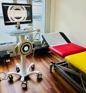
Fig. 1 REMS device at Christian Schneider’s Orthopedic Center Theresie, Munich
Cases from practice
45-year-old professional tennis player with activated OA of the knee including bone marrow edema of the medial femoral condyle. No trauma and no assessment of her bone health to date, as she has not yet reached menopause. No vitamin D supplementation has been given to date. In addition to knee therapy, a bone health screening using REMS was therefore carried out as a supplementary measure. This revealed osteopenia (T –1.3) measured in the femur with a T-score of –0.6 in the lumbar spine. In addition to adjusting her training regimen, vitamin D supplementation is now being administered in accordance with guidelines. Follow-up using REMS is planned in one year, with further diagnostics as needed.
22-year-old professional ice dancer with various recurrent musculoskeletal complaints; currently leading were lumbar spine complaints with bone marrow edema / stress fracture in the vertebral arch LWK 5 both sides incl. surrounding reaction. Part of the diagnostics also included ultrasound-based bone density measurement. This showed a normal lumbar spine but a T-score of –2.3 femoral with an increased 5-year hip fracture risk in the fracture risk assessment. The overall recommendation was core and stability training with partial lumbar orthotic support, kinesiotaping and heat / cold therapy, continuation of eccentric training (regarding knee complaints), as well as selective periradicular injection in the lumbar spine area, magnetic resonance therapy (MBST®) of the spine, and administration of 100.000 I.U. vitamin D3 intramuscularly (every three months). Follow-up using REMS planned after six months and, if necessary, extended diagnostics and training adjustments.
79-year-old female patient with tapping and pressure pain in the upper lumbar spine area without trauma or a history of osteoporosis. Imaging (pelvic and lumbar spine X-ray and lumbar spine MRI) revealed a symphysis-related pubic bone fracture, a degenerative, partially activated spine, and fresh upper plate fractures in L1+2. Osteodensitometry with REMS yielded a T-score of –3.0 (lumbar spine) and –2.9 (femur) with a significantly increased risk of fracture and normal body composition (Fig. 2). Denosumab was administered and a fixed vitamin D substitution was prescribed. The patient is undergoing regular physiotherapy, is mobile in everyday life, and has no complaints. A follow-up examination using REMS is planned in six months and, if necessary, DXA during the course of treatment.
Literature
[1] Kurth A, Kocijan R. Ultraschallbasierte REMS-Technologie in der Diagnostik der Osteoporose und der Beurteilung des Frakturrisikos. Osteologie 2025; 34: 184–191. Doi 10.1055/a-2650-1677
[2] Messina C, Fusco S, Gazzotti S, et al. DXA beyond bone mineral density and the REMS technique: new insights for current radiologists practice. Radiol Med. 2024;129(8):1224-1240. doi:10.1007/s11547-024-01843-6
[3] Pisani P, Conversano F, Muratore M, et al. Fragility Score: a REMS-based indicator for the prediction of incident fragility fractures at 5 years. Aging Clin Exp Res. 2023;35(4):763-773. doi:10.1007/s40520-023-02358-2
[4] Diez-Perez A, Brandi ML, Al-Daghri N, et al. Radiofrequency echographic multi-spectrometry for the in-vivo assessment of bone strength: state of the art-outcomes of an expert consensus meeting organized by the European Society for Clinical and Economic Aspects of Osteoporosis, Osteoarthritis and Musculoskeletal Diseases (ESCEO). Aging Clin Exp Res. 2019;31(10):1375-1389. doi:10.1007/s40520-019-01294-4
[5] Bischoff E, Popova-Belova S, Bischoff F, Kirilov N. Physical Performance of Geriatric Women and Its Impact on Fracture Risk and Bone Mineral Density Assessed with Radiofrequency Echographic Multispectrometry (REMS). Life (Basel). 2024;14(12):1579. Published 2024 Dec 1. doi:10.3390/life14121579
[6] Bobelyak M, Vaculik J, Stepan JJ. Bone mineral density assessment using radiofrequency echographic multispectrometry (REMS) in patients before and after total hip replacement. Osteoporos Int. Published online September 10, 2025. doi:10.1007/s00198-025-07685-w
[7] Borsoi L, Armeni P, Brandi ML. Cost-minimization analysis to support the HTA of Radiofrequency Echographic Multi Spectrometry (REMS) in the diagnosis of osteoporosis. Glob Reg Health Technol Assess. 2023;10:1-11. Published 2023 Feb 6. doi:10.33393/grhta.2023.2492
[8] Kurth A, Optimierung der Versorgung von Patienten mit Fragilitätsfrakturen: Die Rolle von REMS in Tertiären Präventionsprogrammen (z.B. FLS). Osteologie 2025; 34(02): 137-138. doi: 10.1055/s-0045-1804956
Autoren

» Facharzt für Orthopädie mit Zusatzqualifikationen Sportmedizin, Physikalische Therapie, Chirotherapie und Naturheilverfahren.
» Orthopädiezentrum Theresie, München
» U. a. Vorsitzender der Verbandsärzte Deutschland e.V., Vorstandsmitglied in GOTS und Bayerischem Sportärzteverband, Mitglied der medizinischen Kommission des DOSB, Olympiaarzt seit 2006
(Stand 2026)

» Fachärztin für Orthopädie und Unfallchirurgie
» Chief Medical Officer, MBST Global, MedTec Medizintechnik GmbH
» Eberhard Karls Universität Tübingen + AKAD University
(Stand 2026)


