Close Menu
sportärztezeitung
    Facebook YouTube LinkedIn
    • Jetzt Print-Abo sichern!
    YouTube LinkedIn
    sportärztezeitungsportärztezeitung
    • Home
    • Artikel
      • Therapie
      • Training
      • Operation
      • Ernährung
      • Kardiologie
      • Psychologie
    • Health Mediathek
    • Sports Medicine Research
    • Autoren
    • Education
      • Prophylaxis
      • Termine
    • sportlerzeitung
    sportärztezeitung
    Startseite » Thermography plus diagnostic EMG
    Therapie

    Thermography plus diagnostic EMG

    Use in recurrent Achilles tendon symptoms
    Prof. Dr. med. Oliver TobolskiBy Prof. Dr. med. Oliver Tobolski3 Mins Read
    WhatsApp Twitter Email Facebook Telegram Pinterest LinkedIn
    Abb. 1 Wärmebildkamera / FIG. 1 Thermal imaging camera
    Teilen
    WhatsApp Facebook Twitter LinkedIn Email Telegram Copy Link

    Diagnostic thermography has now achieved a relevant status for assessing functional symptoms in sports orthopaedics. Trigger points are readily shown with the help of thermal imaging cameras, especially chronified functional symptoms in the entire axial skeleton, and corresponding therapeutic measures such as shock wave treatments, dry needling, cryotherapy, friction massage or taping can be started and their success monitored in the course of treatment.

    With the infrared thermography we use (FLIR E75 thermal imaging camera, Fig. 1) the camera displays more than 75,000 measured positions with a thermal sensitivity of <0.04 degrees tempe­rature difference, and thus it can reliably demonstrate even small areas of hyperaemia (trigger points).  We can differentiate between active myofascial trigger points which are often extremely painful and frequently weaken the affected muscle, latent myofascial trigger points which are only painful on movement, and associated trigger points which ­develop due to functional disorders of neighbouring muscle groups. More recently, differentiated EMG investi­gations are being used increasingly in conjunction with thermography to identify problems with efferent signals to the musculature in parallel, and to correct these with the help of biofeedback training. Initially thermography was mainly used for muscles close to the vertebral column to identify painful muscular tension in the lumbar and cervical spine and to work specifically on these structures. In the present case the indication for diagnostic thermo­graphy has been extended to Achilles tendon symptoms.

    Case study 

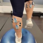
    We were consulted by a 29-year-old [female] patient with recurrent symptoms at the myotendinous junction of the right Achilles tendon, particularly one day after sporting activity (running). Her average training schedule was 40 – 50 km/week, she could not remember having had an accident.  The diagnostic imaging we performed (ultrasound, MRI) and differentiated foot pressure measurements ruled out any structural changes in the tendon and any relevant foot deformities. Only the ultrasonography on the day after spor­ting activity showed peritendinitis in the proximal part of the tendon. The subsequent EMG of the gastrocnemius muscle showed an increased resting tone of the right gastrocnemius. Further functional diagnostics (neuromuscular activation) showed a reduced efferent supply to the medial calf muscles (Figs. 2 & 3). 
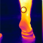
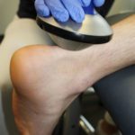
    Abb. 2 Dynamische EMG-Messung des Gastrocnemius (Ruhetonus und Aktivierung erhöht rechts) / Fig. 2 Dynamic EMG measurement of gastrocnemius (resting tone and activation increased on the right)
    Abb. 3 EMG-Messung auf unebenem Untergrund (neuromuskuläre Aktivierung) / Fig. 3 EMG measurements on uneven ground (neuromuscular activation)

    The thermography carried out in pa­rallel impressively showed a painful area in the proximal region of the right Achilles tendon (Figs. 4 & 5). In addition to (focused) shockwave therapy the patient was treated with laser therapy and neuroreflex cryotherapy of the proximal Achilles tendon origin with taping of the muscle and (later) adjunctive biofeedback training of the calf muscles (Figs. 6 – 10).

    Abb. 4 Thermographie mit Nachweis mehrerer Triggerpunkte, sowohl im myotendinösen Übergang als auch im Muskelbauch des M. gastrocnemius , s. Kreise / Fig. 4 Thermography showing several trigger points, both at the myotendinous junction and in the muscle belly of the gastrocnemius muscle, see circles
    Abb. 5 Thermographische Darstellung der Wadenregion in Plantarflexion mit deutlichem Nachweis des Triggerpunktes, s. Kreis / Fig. 5 Thermographic image of the calf in plantar flexion with clear evidence of the trigger point, see circle
    Abb. 6 Neuroreflektorische Kältetherapie (Cryotherapie) der Sehne / Fig. 6 Neuroreflex cryotherapy of the tendon
    Abb. 7 Fokussierte Stoßwellen­behandlung der Triggerpunkte / Fig. 7 Focused shockwave treatment of the trigger points
    Abb. 8 Laserbehandlung der Achillessehnen / Fig. 8 Laser treatment of the Achilles tendons
    Abb. 9 Tape-Anlage / Fig. 9 Applying tape
    Abb. 10 EMG-gesteuertes Biofeedbacktraining der Wadenmuskulatur / Fig. 10 EMG-guided biofeedback training of the calf muscles

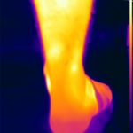
    The follow-up examinations showed a marked reduction in trigger point formation over the proximal Achilles tendon, even after the first treatment session. After two sessions of shockwave therapy plus taping and two sessions of biofeedback training the patient was clinically 100% symptom-free (Figs. 11 – 13).

    Abb. 11 Rückläufige Triggerpunkt­bildung im myotendinösen Übergang (nach 1. Behandlung) / Fig. 11 Regression of trigger point formation at the myotendinous junction (after 1 treatment)
    Abb. 12 Fast vollständige Rück­bildung des Triggerpunktes in der Thermo­graphie (nach 3. Behandlung) / Fig. 12 Virtually complete regression of the trigger point in the thermography (after 3rd treatment)
    Abb. 13 Vollständige Rückbildung des Triggerpunktes auch in Belastung (Plantarflexion) / Fig. 13 Complete regression of the trigger point, also on exertion (plantar flexion)

    Summary

    Thermography impressively helps diagnose muscular trigger points and moni­tor successful treatment, particularly of functional symptoms, including those in the tendinous origins distant from the spine. Secondary muscle dysfunction is demonstrated by EMG and treated ­using EMG-based biofeedback therapy.

    Autoren

    Prof. Dr. med. Oliver Tobolski

    ist Leiter von Ortho4Sport in Köln. Er ist Facharzt für Chirurgie mit Zusatzbezeichnungen Sportmedizin und Chirotherapie. Außerdem ist Professor Tobolski Verbandsarzt des Tennisverbandes Mittelrhein sowie ATP-Turnierarzt.

    INT 23
    Share. WhatsApp Facebook Twitter LinkedIn Telegram Email
    Previous ArticleACP-Therapie
    Next Article The burden of hamstring injuries in professional footballers

    Weitere Artikel aus dieser Rubrik

    Therapie

    sports.medicine.newspaper 2025

    By Angie Sarka
    Therapie

    Neues aus der Arthroseforschung

    By Dr. rer. nat. Wolfgang Feil
    Therapie

    Early Postoperative Combined Physical Therapy Following Hamstring Tendon Refixation

    By Dr. med. Tobias Würfel , Peter Stiller

    Neueste Beiträge

    sports.medicine.newspaper 2025

    Therapie Angie SarkaBy Angie Sarka

    Neues aus der Arthroseforschung

    Therapie Dr. rer. nat. Wolfgang FeilBy Dr. rer. nat. Wolfgang Feil

    Early Postoperative Combined Physical Therapy Following Hamstring Tendon Refixation

    Therapie Dr. med. Tobias Würfel , Peter StillerBy Dr. med. Tobias Würfel , Peter Stiller

    Achilles tendonopathy

    Therapie Dr. med. Henning Ott, Dr. med. Julia Walter , Larissa TheisBy Dr. med. Henning Ott, Dr. med. Julia Walter , Larissa Theis

    Collagène

    Ernährung Miguel Florido, MDBy Miguel Florido, MD

    Anstehende Veranstaltungen

    März 15
    15. März um 09:00 - 9. November um 17:00

    Workshops 2025 – Stoßwellen- /Lasertherapie & alternierende Verfahren

    Mai 24
    Ganztägig

    Kongress für Ernährungstherapie im Kontext Sportmedizin & Prophylaxe

    Nov. 15
    08:00 - 17:00

    15 .Symposium der sportärztezeitung

    Kalender anzeigen

    Newsletter abonnieren

    Erhalten Sie aktuelle Informationen zu den neuesten Artikeln, Studien und Veranstaltungen.

    Sportmedizin für Ärzte, Therapeuten und Trainer

    YouTube LinkedIn
    Rubriken
    • Therapie
    • Training
    • Ernährung
    • Operation
    • Kardiologie
    • Applikation
    • Psychologie

    Jetzt zum Newsletter anmelden

    Mit unserem Newsletter keine Beiträge und Neuigkeiten mehr verpassen.

    Copyright ©thesportGroup GmbH
    • Impressum
    • Cookie Einstellungen
    • Datenschutzerklärung

    Type above and press Enter to search. Press Esc to cancel.