Osteitis pubis, a clinical picture that is frequently described in sports medicine and gynaecology, is usually complex for the practitioner/therapist. Exercise-induced groin pain often limits training and is very painfully tedious for athletes and active people.
One patient group consists of young football players. The onset of inflammation in the pubic rami is very often caused by muscular imbalance that develops due to one-sided training and asymmetrical loading patterns. This is compounded by hard running surfaces e.g. artificial turf pitches, and a lack of mobility and length of the muscles close to the pelvis. In a large number of treated cases, premature and excessively intensive increases in exercise lead to recurrence and protracted downtimes. Another patient group consists of postpartum women. During the pregnancy hormones cause loosening of the pelvic ligamentous structures. This laxity in the last trimester of the pregnancy provides for widening of the birth canal, thus easing the delivery process. In a physiological situation these ligamentous structures tighten again postpartum. Any disorder of tightening of the ligamentous apparatus can lead to severe protracted pain and massive restrictions in everyday living for the women affected. A history of recalcitrant and protracted pain is commonly typical in both patient groups.
Diagnostic imaging by MRI or ultrasound examination, if necessary, can confirm the diagnosis and distinguish between a number of differential diagnoses (e.g. torn adductor(s), rectus abdominis injury, inguinal hernia). A prominent feature in an MRI scan is bone marrow oedema, usually encountered bilaterally, in the pubic regions close to the symphysis. Oedematous spread is usually asymmetrical and mostly correlates with the side with more conspicuous symptoms. The so-called secondary cleft sign, the formation of an asymmetrical fissure, can often be seen within the symphysis. In many cases, poor muscular control of the local stabilisers of the lumbar spine and the hip joint as well as hypertonia, e.g. of the iliopsoas muscle, can be observed. The effects of using extracorporeal shockwave therapy to treat musculoskeletal tissue have been compiled comprehensively in a recent systematic review [1]. When treating bone, extracorporeal shockwave treatment causes activation of the stem cells, the osteoblasts. Hence radial extracorporeal shockwave therapy should be included in the holistic treatment regimen for this indication.
Showing the muscle activity with the help of electromyography (EMG) is very helpful for recognising neuromuscular activity patterns which are then applied specifically in biofeedback training. This enables the therapist and patient to objectify and feel how the specific target musculature is optimally trained and activated. Since its start a few years ago, patients can access training plans based on an app to accompany the therapy process and promote the patient’s autonomous training. This way the physiotherapist/trainer can draw up the individual exercise programme and forward it with an app to the patient who, in turn, documents his/her training including the course of the pain.
Case Report
The [female] patient, 32, athletic with persisting and chronic progressive pain in the region of the symphysis with increased radiation into the left groin presented at the practice. The pain began in the last third of her pregnancy, but these pains have now been constant for 1.5 years and limit all activities of daily life. Over the course of time she had tried several classic physiotherapy approaches and treatment, all of which, however, resulted in brief improvement at most. As soon as she increased the intensity of her activity and training in the past the pain reoccurred very severely. For his diagnosis the attending physician had ordered another MRI examination which, compared with the previous examination, showed spread of the bone marrow oedema on both sides, and the left ramus of the pubic bone was clearly more conspicuous. During the gait analysis at the baseline examination the patient showed considerably shortened step length on the left and an increase in the stride width. During the standing examination the lordosis of the lumbar spine was seen to be very pronounced, which was associated with poor activity of the anterior musculature. Her left-sided pain could be provoked by her standing on one leg. The Thomas test was conspicuous on both sides. The manual therapy examination of the lumbar spine and the hip joint was inconspicuous and was not connected with her pain.
Treatment Protocol
DAY 1: TREATMENT 1
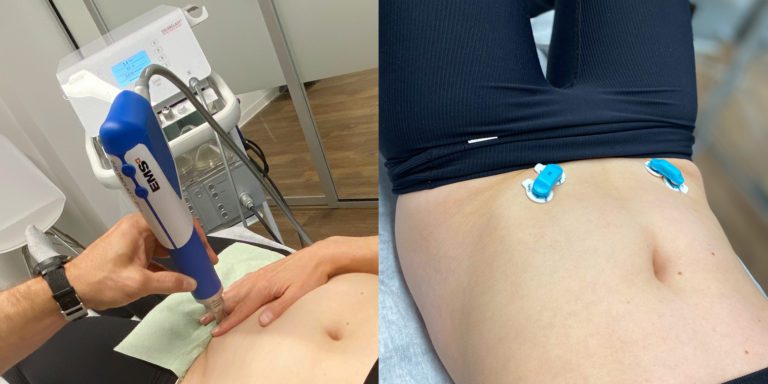
- Radial ESWT (Electro Medical System, Swiss Dolorclast Evoblue, Nyon – 25 Hz; 1.4 bar; 1.5 cm handpiece; 3000 impulses targeted at the region of the bone marrow oedema)
- Myofascial treatment of the iliopsoas muscle.
 Fig. 1 Treatment of bone marrow oedema/pubic bone. Handpiece is held/guided by the patient’s fingers.
DAY 5: TREATMENT 2
- radial ESWT (25 Hz; 1.8 bar 4500 impulses)
- Myofascial treatment of the iliopsoas muscle.
- Training programme drawn up (App: Lanista Athlet, MP Sports Coaching & Consulting GmbH, Munich) – bridging on both sides, squats, segmental pelvic movements sitting.
DAY 9: TREATMENT 3
- Pain-free in everyday living since second treatment; step length and stride width normal
- Radial ESWT (25 Hz; 2.6 bar 5000 impulses)
- Biofeedback Training (EMG System, Menios GmbH, Ratingen) local stabilisers (multifidi, int./ext. obliques).
- Expanded training programme – activation of the local stabilisers incl. pelvic floor in diverse functional starting positions.

DAY 21: TREATMENT 4
- Radial ESWT (25 Hz; 2.7 bar 5000 impulses)
- Biofeedback training of the local stabilisers with dynamic asymmetric exercises (single-legged squats, standing balance exercise, side lunge)
- Expanded training programme – asymmetrical exercises, some dynamic
- MRI check-up before treatment 5: ”Complete regression of the bone marrow oedema in the pubic bone on both sides, no evidence of any other pathological conditions in the region examined“.
DAY 48: TREATMENT 5
- Radial ESWT (25 Hz; 2.7 bar 4500 impulses)
- Biofeedback training of local stabilisers when jumping (two-legged, one-legged short distance)
- Exercise programme drawn up with the training objective of 30 min. pain-free jogging
Summary
With the help of radial shockwave therapy the patient was quickly relieved of her chronic progressive pain. Thus, the new radiology appointment showed complete resolution of the bone marrow oedema. The possibility of using EMG in the biofeedback training resulted in effective and targeted training. By using digital training planning the patient was able to work on her muscular deficits autonomously under ongoing checks by the physiotherapists. The patient’s motivation and compliance were positively supported by these digital checks on her training results. However, the research carried out for this article has also showed that there are as yet no generally valid training protocols for this indication, and that training very often depends on the practical experience of the therapist/trainer. Further sports/physiotherapy studies/training protocols would be helpful for this.
Literatur
[1] Wuerfel, T.; Schmitz, C.; Jokinen, L.L.J. The Effects of the Exposure of Musculoskeletal Tissue to Extracorporeal Shock Waves. Biomedicines 2022, 10, 1084. https://doi.org/10.3390/biomedicines10051084
Autoren
ist Physiotherapeut, OMPT-DVMT | HP-PT und international anerkannter Manualtherapeut (IFOMPT) mit eigener Praxis in Landshut (TMPHYSIO Mühleninsel).


